2D Echo Test in Nagpur
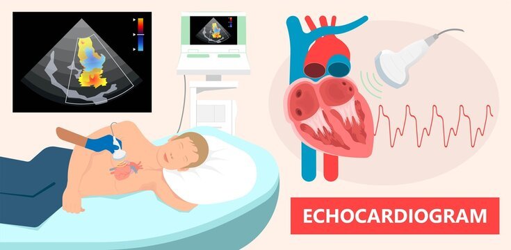
Know everything about the 2D Echo test?
Have you lately felt out of breath while climbing stairs to your office on the second floor? Otherwise you felt low on stamina amid your weekend football match along with your friends, something that you simply used to appreciate for hours. These are the signs that your heart needs more attention and care.
Our heart pumps blood, and the vascular framework takes it to all the organs. Together they shape the cardiovascular framework. And to do its work appropriately, all your heart requires is normal exercise, a balanced and nutritious diet, and administration of unnecessary stress.
In any case, as your age advances, you wish to incorporate one fundamental thing in your healthy heart lifestyle – 2D echocardiography – to guarantee your heart wellbeing from time to time.
What is 2D Echo Test?
A two-dimensional Echocardiogram or 2D Echo test is a demonstrative test that employs ultrasound waves to evaluate the working of the heart. When these waves hit the organ structure’s interior, they echo back and produce moving pictures of the heart and valves on the computer screen.
The image hence formed is known as Echocardiogram. This is often utilized to evaluate any breakdown or harm related with the heart tissue or valves like clots, blockage, inherent heart defects, and issues like coronary artery illnesses.
It is a non-invasive method with no side effects. However, one may feel marginally awkward during transducer arrangement because it puts pressure on the heart.
How is 2D ECHO performed?
A 2D ECHO exam is performed using a portable transducer unit that can be carried to the patient’s bedside. The 2D echo procedure for this test depends on the type of test you are running.
Transthoracic echocardiography:
In this procedure, ultrasound-emitting transducers are placed at various locations on the chest to obtain instantaneous images of the heart.
Stress echocardiography:
It runs in her two stages. First arrange, a resting echocardiogram is taken with the person lying on the cleared outside with the left arm expanded. Within the second stage, he is constrained to do moderate work out, such as running on a treadmill on an incline, for 5-15 minutes, depending on wellness level and age. During this procedure, a blood pressure sleeve is connected to the upper arm and terminals are joined to the chest and legs to always screen blood weight and an EKG. In uncommon cases, chest inconvenience, tipsiness, or migraine may happen amid this strategy. Drugs such as sildenafil can influence the coming of this test.
Transesophageal echocardiography:
In this procedure of 2D echo test, a transducer is attached to a probe that is inserted into the throat and emits ultrasound waves that bounce off the heart to obtain different views of the heart. A drug may be injected to help you relax during the exam, and a spray or gel will numb the back of your mouth before the probe is inserted. You may experience a sore throat after completing this test and may not eat or drink until 30 to 60 minutes after the test.
Why is 2D ECHO performed?
2D ECHO tests are done mainly to detect various heart diseases and problems. A doctor recommends checking out your Health 2D ECHO scan to better understand the following:
Size and shape of Heart
The heart can enlarge due to high blood pressure (BP), or the heart’s lower chambers, the ventricles, can enlarge for various reasons and lead to ineffective pumping.
Sac surrounding the heart
In some cases, a buildup of fluid can occur inside the pericardium and cause compression of the heart leading to signs and symptoms, such as a sharp drop in blood pressure, low heart sound, shortness of breath, weakness and cough.
Congenital heart disease
Newborn heart problems, such as holes in the heart wall, can be detected with 2D ECHO.
Heart valves
The 2D ECHO test can be used to correlate symptoms of valvular dysfunction, such as fatigue, leg swelling, and changes in heart sound during auscultation.
Blood clots
Patients with an ongoing stroke or a history of stroke are recommended to have this test performed to detect the location and extent of damage to heart tissue.
Cardiac muscle
Heart attacks can weaken the heart muscle, which further reduces the blood supply to the heart. This weakened muscle may not contract as effectively as it did before the heart attack.
How do you plan for the 2D Echo test?
- For TTE, no particular arrangement is required earlier to the test, making it less demanding and more profitable than other tests when it comes to planning.
- For stress echocardiography, the person must not eat or drink anything for three hours some time during the test. Free and comfortable clothing must be worn. Too, it is vital to educate the doctor on the off chance that you’re taking solutions such as sildenafil (Viagra).
- TEE is for the most part done for hospitalized patients. Fasting for a few hours is required some time in this test, and the precise length for fasting is educated by the treating doctor. The persistent must educate the doctor approximately sensitivities to any solutions. Dentures must be evacuated earlier to the test.
If you are in Nagpur and are facing some problem with your heart, reach out to the best cardiologist in Nagpur that is Dr. Chetan Rathi. Check out right away.
Our Medical Services

ECG
An electrocardiogram (ECG) is one of the only and speediest tests utilized to survey the heart. Anodes (small, plastic patches that stick to the skin) are set at certain spots on the chest, arms, and legs.

Best Cardiologist in Nagpur
Introducing Dr. Chetan Rathi, a distinguished Cardiologist in Nagpur, whose eminence transcends the realm of medical proficiency.

Holter Monitoring
Holter monitoring measures your heart activity over an extended period, usually between 24 and 48 hours. Basically, a Holter Monitoring is a portable device which records the heart’s electrical signals.

BP Monitoring
Each time your heart beats, it pumps blood into your arteries. A blood pressure measurement may be a test that measures the force (pressure) in your arteries as your heart pumps.

Coronary Angiography
Coronary angiography diagnoses and evaluates coronary artery blockages. Contrast dye is injected into arteries, enabling X-ray imaging to visualize blood flow and identify narrowing or blockages.
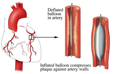
Coronary Angioplasty
Coronary angioplasty is a minimally invasive technique of abdominal artery angioplasty, which is used to treat coronary arteries that are obstructed or constricted and it is the most appropriate technique used by doctors for the treatment.
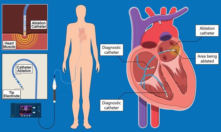
Electrophysiology Study
An Electrophysiology Study (EP study) is a test utilized to assess the heart’s electrical framework and check for abnormal heart rhythms. The natural electrical impulses coordinate the contractions of different parts of the heart.
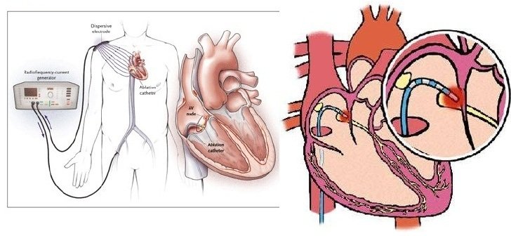
Radiofrequency Ablation
Radiofrequency Ablation (RFA) is a minimally invasive medical procedure that uses high-frequency electrical currents to generate heat, effectively destroying abnormal tissue or cells.
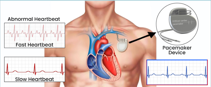
Pacemaker Implantation
Pacemakers are medical devices designed to support and regulate the electrical system of the heart, ensuring it functions properly. This medical procedure entails the insertion of a small device into the chest region.

ICD Implantation
An Implantable Cardioverter-Defibrillator (ICD) is a life-saving device that plays a crucial role in monitoring and regulating heart rhythms. It consists of a pulse generator and leads implanted in the heart.

CRT_P & CRT-D Implantation
CRT implantation is a process in which technological instruments known as CRT-P and CRT-D where p stands for pacemaker and d stands for defibrillator.

Valvuloplasty
A balloon mitral valvuloplasty is a process to extend a restricted heart valve and improve blood flow. The heart valves handle how blood drives through the heart.
Our Achievements in Numbers
Happy Patients
Years of experience
Specialisations
Hospital Associations
Awards & Recognition
Patient Testimonials
Dr Chetan Rathi sir is good cardiologist. I visited him many times with my family and friends for consult about issues related with cardiology.
Our Videos


Our Blog
 What are the Reasons for Heart Attack in Women and How to Prevent Them
Women’s heart attacks can be triggered by various reasons including smoking, high blood pressure, diabetes, obesity, and stress among others. The measures include doing regular exercises and other physical activities, having a balanced diet, non-use of tobacco products, stress management, and consulting a doctor for any health complications. What is Heart Disease? These are illnesses… Continue reading What are the Reasons for Heart Attack in Women and How to Prevent Them
Read More Lifestyle Changes After Angioplasty
Angioplasty is a frequently utilized technique involving the use of a small balloon to open blocked or narrowed coronary arteries. After angioplasty, the patient has to change his or her lifestyle to avoid potential risks to the heart in the future. What is an Angioplasty? Angioplasty is a specific surgical technique in which a balloon… Continue reading Lifestyle Changes After Angioplasty
Read More Difference Between ECG vs ECHO
Have you ever wondered about the difference between an ECG and an ECHO? If you’ve had a heart checkup or are curious about heart health, these two tests might sound familiar. While both focus on your heart, they do very different things. Let’s break down what sets them apart. ECGs and Echocardiograms Electrocardiograms (ECG or… Continue reading Difference Between ECG vs ECHO
Read More


