EP Study Test in Nagpur
If a racing heart, dizziness or fainting without explanation is the problem you or a family member is experiencing, your doctor may suggest that you undergo an EP study for heart health. In case you decide to get any medical treatment, it is natural to feel scared. However, if you are told about it in detail and plainly, you may feel less frightened. Teachers always suggest reading before the lesson is taught. Similarly, this book will explain to you what the EP study test in Nagpur involves. In fact, it is like having someone at your side during the whole process.
Below you can find detailed steps for how the EP study test is done in Nagpur including the facility where the test is done.
What is EP study?
An Electrophysiology Study (EP study) is a test utilized to assess the heart’s electrical framework and check for abnormal heart rhythms. The natural electrical impulses coordinate the contractions of different parts of the heart. This helps maintain normal blood circulation. This movement of the heart produces the heartbeat. This is done so the heart can be evaluated and its damaging causes can be found. It may also be done to help evaluate how your body responds to some medicines.
During an EP study, a heart rhythm specialist or an electrophysiologist can also map the propagation of electrical impulses from the heart with each beat. This may be done to help find the source of the irregular heartbeat.
What are the Procedure of EP Study Test
The whole process of an EP study is carried out in a special room called a Cath Lab. Here’s the procedure in a simple step-by-step guide for you:
1. Preparation
The medical team will shave the part of your body where the tubes are supposed to be inserted, which is usually your groin. They will put you on a monitor to check your heart rate and blood pressure continuously. Besides, you will get some medicine through an IV to help you relax.
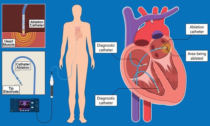
2. Catheter Insertion
The doctor will first administer a local anaesthetic injection so that the skin in your groin is numbed. After that, he or she will insert thin, flexible tubes, known as catheters, into a vessel of the bloodstream and guide them gently into your heart.
3. Electrode Placement
Electrodes are mini metal portions placed at the end of catheters. Using fluoroscopy, the doctor is able to see the catheters’ journey through the heart and get them to the right places in the heart chambers to pick up the electrical signals directly at the source.
4. Electrical Stimulation
Electrically stimulating the heart is the method by which the doctor sends small, safe electrical impulses through the catheters so that the heart can be made to beat at different speeds. In this way of conducting the test, the doctor will be able to show the abnormal heart rhythm, its beginning, and how it is moving.
5. Data Collection
During the stimulation, special computers document in real time the electrical activity of the heart. Based on the analysis of these recordings, the doctor can pinpoint the exact locations of the aberrant electrical signals.
6. Diagnosis and Treatment
Once the problem area is found, the doctor makes a final diagnosis. In many cases, they can treat the issue right then and there using a procedure called ablation, which uses heat or cold to neutralize the tiny spot causing the trouble.
What are the Benefits of an EP Study test?
The aims of conducting EP studies are to gain insight into the function of each component of the conduction system, to identify the mechanism and precise focus of arrhythmia, to perform risk stratification, and to perform ablation of the impaired circuit. to determine the need for treatment or therapy. This article provides an overview of anatomy and physiology, common cardiac indications, and the clinical importance of EP research. On average, the electrophysiology study cost of a basic EP study without ablation could range from ₹50,000 to ₹1,50,000 or more. If ablation or other interventions are required, the cost would increase significantly.
Why is EP Study Test done?
Doctors order this test to find the root cause of abnormal heart rhythms, also known as arrhythmias. If your heart beats too fast, too slow, or irregularly, standard tests might not catch it if it happens randomly. An EP study allows a heart specialist to safely trigger the irregular rhythm in a controlled lab, find out exactly where the glitch is, and decide the best way to fix it.
What are the Risks of an EP study test?
In most cases, this procedure is quite safe. Potential risks may include things such as arrhythmia. An arrhythmia may occur during the EP examination and cause dizziness. In this case, your doctor may give your heart an electric shock to get it back to normal. A heart attack (myocardial infarction), stroke, or damage to a heart valve can also be fatal.
In some cases, a blood clot forms at the tip of the catheter and breaks off, blocking the blood vessel. Your doctor may prescribe drugs to prevent blood clots. Infection, bleeding, and bruising at the catheter insertion site (groin, arm, or neck) may also occur. Doctors and nurses can help avoid these problems.
- Bleeding or infection.
- Bleeding around the heart caused by damage to the heart tissue.
- Damage to the heart valves or blood vessels.
- Damage to the heart’s electrical system, which could require a pacemaker to correct.
- Blood clots in the legs or lungs.
- Heart attack.
- Stroke
What you can expect from EP Study Test
Everything you need to know before, during, and after taking the EP Study Test.
Before an EP Study Test
- You will change into a hospital gown and sign a consent form.
- The nurse will start an IV line in your arm for fluids and medicines.
- The medical team will double-check your identity and the details of your test.
During an EP study Test
- In hospitals and clinics, doctors and nurses perform EP tests in rooms with special testing equipment. This room may be called the “Electrophysiology Laboratory” or the “EP Laboratory” or some call it a catheterization lab. During testing:
- If the type and location of the arrhythmia have been identified and appropriate therapy determined, cardiac ablation or insertion of a cardiac pacemaker or his ICD can be performed during or shortly after the EP study.
- A nurse will plug her IV (infusion line) into your arm. You will be given drugs to help you relax (sedatives). However, during the test I am awake and able to follow instructions.
- It usually occurs in the groin, but can also occur in the arms and neck.
- An injection of local anesthetic is given to numb the area. A doctor inserts a needle through the skin and into a blood vessel.
- Your doctor sends small electrical pulses through the catheter to make your heart beat at different rates. You may feel your heart beat faster and faster.
- Electrical signals produced by the heart are picked up and recorded by a special catheter. This is called cardiac mapping and allows doctors to pinpoint the cause of an abnormal heart rhythm.
- Your doctor will remove the catheter and IV line. The nurse will apply pressure to the injection site to stop the bleeding. The test usually lasts 1-4 hours.
Key Parts of Electrophysiology Study
An electrophysiology (EP) study involves several key parts that help doctors understand and manage heart rhythm issues:
Catheter Insertion
Thin, flexible tubes called catheters are carefully inserted into blood vessels, typically in the groin or neck area, and guided to the heart.
Mapping and Recording
Catheters record the heart’s electrical signals, creating a map that helps locate abnormal areas or irregular heart rhythms.
Provocation Techniques
Using the catheters, doctors can trigger or provoke abnormal heart rhythms to understand how and where they originate.
What happen after the Procedure EP Study?
You will be moved to the recovery room for 1 to 3 hours. In this time:
- Lie still for as long as the nurse tells you to. Be sure to keep the arm or leg used for the test straight.
- Your nurse will check for bleeding or swelling at the puncture site.
- Before you leave, you will be told what to do at home. Follow the instructions your nurse or doctor has given you, including taking any new medicines that have been prescribed for you. Most people can start eating and taking medicine within 4 to 6 hours after the test. Most are able to do their usual daily activities the day after the test. Do not drive for at least 24 hours.
- The needle puncture site may be painful for several days. A small bruise at the puncture site is normal. If the site begins to bleed, lie flat and press firmly on it. Have someone call your doctor or EP lab.
Till date patients of Nagpur were referred to metropolitan cities like Mumbai or Hyderabad for this treatment or senior EP specialists used to visit Nagpur to treat patients. With full time accessibility of Dr. Chetan Rathi at Nagpur, 1st EP specialist of not only Nagpur but whole Vidarbha locale, patients can indeed profit from this facility at Nagpur.
Our Medical Services

ECG
An electrocardiogram (ECG) is one of the only and speediest tests utilized to survey the heart. Anodes (small, plastic patches that stick to the skin) are set at certain spots on the chest, arms, and legs.
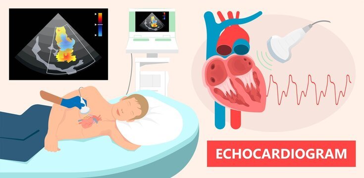
2D Echo
A two-dimensional Echocardiogram or 2D Echo test is a demonstrative test that employs ultrasound waves to evaluate the working of the heart.

Holter Monitoring
Holter monitoring measures your heart activity over an extended period, usually between 24 and 48 hours. Basically, a Holter Monitoring is a portable device which records the heart’s electrical signals.

BP Monitoring
Each time your heart beats, it pumps blood into your arteries. A blood pressure measurement may be a test that measures the force (pressure) in your arteries as your heart pumps.

Coronary Angiography
Coronary angiography diagnoses and evaluates coronary artery blockages. Contrast dye is injected into arteries, enabling X-ray imaging to visualize blood flow and identify narrowing or blockages.
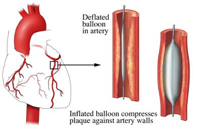
Coronary Angioplasty
Coronary angioplasty is a minimally invasive technique of abdominal artery angioplasty, which is used to treat coronary arteries that are obstructed or constricted and it is the most appropriate technique used by doctors for the treatment.

Best Cardiologist in Nagpur
Introducing Dr. Chetan Rathi, a distinguished Cardiologist in Nagpur, whose eminence transcends the realm of medical proficiency.
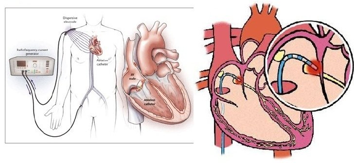
Radiofrequency Ablation
Radiofrequency Ablation (RFA) is a minimally invasive medical procedure that uses high-frequency electrical currents to generate heat, effectively destroying abnormal tissue or cells.
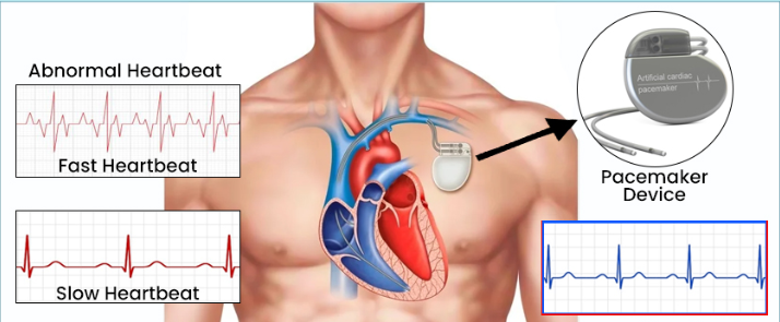
Pacemaker Implantation
Pacemakers are medical devices designed to support and regulate the electrical system of the heart, ensuring it functions properly. This medical procedure entails the insertion of a small device into the chest region.

ICD Implantation
An Implantable Cardioverter-Defibrillator (ICD) is a life-saving device that plays a crucial role in monitoring and regulating heart rhythms. It consists of a pulse generator and leads implanted in the heart.

CRT_P & CRT-D Implantation
CRT implantation is a process in which technological instruments known as CRT-P and CRT-D where p stands for pacemaker and d stands for defibrillator.

Valvuloplasty
A balloon mitral valvuloplasty is a process to extend a restricted heart valve and improve blood flow. The heart valves handle how blood drives through the heart.
Our Achievements in Numbers
Happy Patients
Years of Overall Experience
Specialisations
Hospital Associations
Awards & Recognition
Patient Testimonials
 Girish Kale
Treatment was superb and counselling to patient is very good. My uncle feels very comfortable while getting treated by Dr Rathi.
 Swapnil Chouhan
Doctor Chetan Rathi sir is a very nice and supportive. He explains everything very clearly. The hospital staff is very neat, clean and disciplined. I have had a very good experience here.
 Kuldeep Kapoor
Dr Chetan Rathi Sir is very Polite in nature. He gives full information to Patient regarding their illness. He gives ample of time to Patient in his Chamber.
 Shamod Jadhao
I visited Dr Rathi's clinic as I was having chest pain and palpitation. One of my friend suggested me. He was so calm and friendly listened to my concern very carefully.
Dr Chetan Rathi sir is good cardiologist. I visited him many times with my family and friends for consult about issues related with cardiology.
Our Videos


Our Blog
 Structure and Function of Heart Chambers Explained
The heart is the most hardworking part of your body. It is a muscle that acts like a pump to send blood everywhere, from your head to your toes. To carry out this work, your heart has a very special design. Let’s see how the heart chambers help keep your body healthy. What are the… Continue reading Structure and Function of Heart Chambers Explained
Read More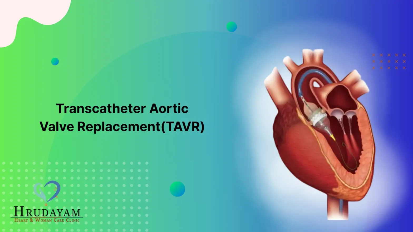 Transcatheter Aortic Valve Replacement (TAVR): Procedure, Benefits & Complications
Your heart functions like a constantly working pump. It has small doors called valves that are responsible for making sure the blood flows in the right direction. Sometimes, these valve doors become stiff and stop opening properly. A replacement procedure named TAVR can come to your rescue in such a situation. Let us find out… Continue reading Transcatheter Aortic Valve Replacement (TAVR): Procedure, Benefits & Complications
Read More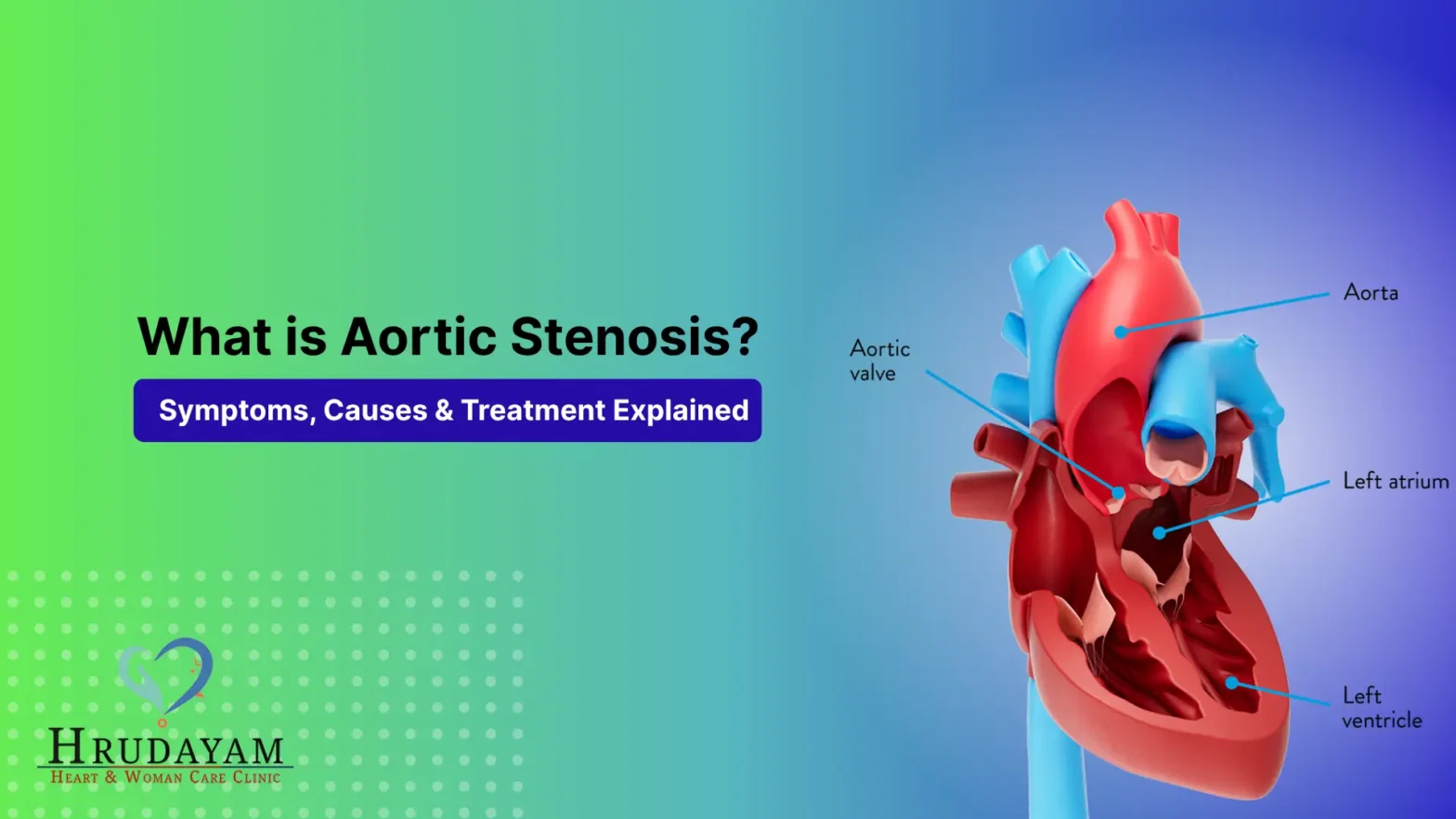 Aortic Stenosis? Symptoms, Causes & Treatment Explained
The heart is like the engine of our body. It circulates blood to every part of us to sustain life. However, one of the “doors” in the heart, also known as a valve, might become narrow. This is referred to as aortic stenosis. When this occurs, the heart has to strain itself significantly in order… Continue reading Aortic Stenosis? Symptoms, Causes & Treatment Explained
Read More